Preface

This purpose of this page is to assist the reader in beginning to think about heart rhythms and heart blocks in the context of the heart's conduction system, and assumes a pre-existing basic competence in rhythm identification. When a clinician begins thinking about heart rhythms and heart blocks in the context of the conduction system instead of rote memorization of ECG patterns, the clinician will become better able to identify rhythms quickly and accurately. The clinician will also become better able to identify more complicated rhythms that do not fit as cleanly into the ECG patterns commonly taught in schools and textbooks.

This page does not provide definitions of rhythms and blocks. The focus here is only to help clinicians already familiar with the definitions to begin to understand why rhythms and blocks manifest as they do on an ECG strip. To that end, we will use the words "always," and "never," as frequently as possible. "Always" & "never" can be scary words to use, because they can make statements vulnerable to attack from small and irrelevant edge cases, but these words are also very powerful; by identifying things that are "always" true, and "never" true, you can greatly improve your ability to quickly and accurately assess different situations.

Some useful tips to aid in rhythm identification:

Atrial Rhythms

Remember that in normal hearts the AV node is the only pathway between the atria and the ventricles. This means that the atria and ventricles are electrically-independent, and only communicate through the AV node. This communication can go forwards, or backwards. It can also be slowed (as in a 1° AV-block) or stopped. If stopped, it can be stopped intermittently (as in a 2° AV-block), or completely (as in a 3° AV-block).

Most of the time the atria and the ventricles work in synchrony and harmony, so we think of the heart as having one rhythm. But when communication at the AV node is impaired or cut off, the atria may keep beating to their own rhythm, while the ventricles begin beating to a separate rhythm, either their own (a ventricular rhythm), or the rhythm of the AV-node (a junctional rhythm). One thing to keep in mind when identifying a rhythm, is that with the exception of some lethal arrhythmias, junctional and ventricular pacemakers emit regular impulses, producing regular rhythms.

With this in mind, let us examine the following rhythm:
ECG strip with an irregular narrow-complex rhythm & P-waves equidistant before all QRS complexes

It is narrow-complex, which rules out ventricular rhythms. It is irregular which rules out both junctional and ventricular rhythms (again, with the exception of some lethal irregular ventricular arrhythmias, which this is clearly not). Therefore, even if there were no visible P-waves, we can be confident that this rhythm is atrial in origin. Sinus arrhythmias like this one are often mistaken for A-fib when the P-waves are small and difficult to see, especially if there's some artifact. When in doubt, lewis leads can be very helpful in identifying difficult-to-see P-waves.

Junctional Rhythms

There are few ECG features that are unique to junctional rhythms, so identification of junctional rhythms should focus on ruling out atrial and ventricular rhythms. When attempting to confirm or rule out a junctional rhythm, remember that junctional rhythms are always perfectly regular. No exceptions! But note that atrial and ventricular rhythms can also be perfectly regular. Junctional rhythms can also be either narrow or wide-complex, so we need understanding of more variables to help us confidently identify junctional rhythms.

As a side-note, we should probably mention that junctional rhythms can appear irregular if they are interspersed with premature atrial and ventricular beats, or if they are transitioning back-and-forth between an atrial or ventricular rhythm and the junctional rhythm, but remember, ignoring rhythm transitions and premature beats, the junctional rhythm itself will always be perfectly regular.

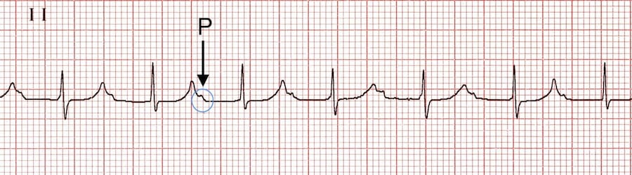
P-R intervals in junctional rhythms are always less than 0.12 seconds (if the P-wave even exists). Please don't merely memorize this, understand why! The P-R interval must be less than 0.12 in a junctional rhythm because it always takes more than 0.12 seconds for an electrical impulse to travel from the atria to the ventricles through the AV node, so if the space between the P wave and the QRS complex is less than 0.12, then the impulse causing the ventricular depolarization can't be coming from the atria! Ergo, it must either be a ventricular rhythm or a junctional rhythm. A narrow-complex would allow you to further rule out ventricular rhythms, leaving only a junctional rhythm as a possibility.

But if the depolarization impulse is not coming from the atria, then why is there often still a P-wave associated with the QRS complex? This is often because of something called "retrograde depolarization." Signals from the junction and ventricles can go backwards up the conduction system and depolarize the atrium, causing a P-wave. P-waves caused by this retrograde depolirization are often (but not always) inverted in the inferior leads like lead II because the electricity is travelling in the opposite direction (bottom-to-top instead of top-to-bottom) as the atrium depolarizes, resulting in an upside-down P-wave. It's important to understand that depolarization of the AV node and conduction system itself doesn't register at all on the ECG strip; the fibers of the conduction system are simply not enough tissue. You don't see anything on the ECG strip until the electricity exits the conduction system and starts depolarizing muscle tissue. There's a lot more cells in the muscle than there are in the conduction system, hence a lot more electricity moving around. If an impulse originates in the junction and reaches the atrium before the ventricles, the P-wave will appear in front of the QRS complex. If that same impulse arrives at the ventricles before it arrives at the atrium, the P-wave will appear either after the QRS complex, or it will be buried inside the QRS complex if the QRS complex is wide enough to eat the P-wave.

Don't use inverted P-waves to identify junctional rhythms. Inverted (upside-down) P waves are neither specific nor 100% sensitive to junctional rhythms. Inverted P-waves (in the inferior leads) are often produced by retrograde depolarization from both junctional and ventricular rhythms, but retrograde depolarization can also produce upright P-waves, and even some impulses originating in the atrium (for example an ectopic focus low in the atrium) can produce upside-down P-waves. Inverted P-waves are a good clue that you may be looking at a junctional rhythm, but they can't be used to confirm or rule out anything. On the other hand, when PR intervals are abnormally short (less than 0.12), or the P-waves are consistently in the "wrong" location (after the QRS or buried inside the QRS), and the number of P-waves matches the number of QRS complexes, then that is conclusive evidence that you're looking at either a junctional or ventricular rhythm. And if the rhythm is narrow-complex and has a PR interval less than 0.12 (or is narrow-complex and matches the conditions described above), then that's conclusive evidence that you're looking at a junctional rhythm.

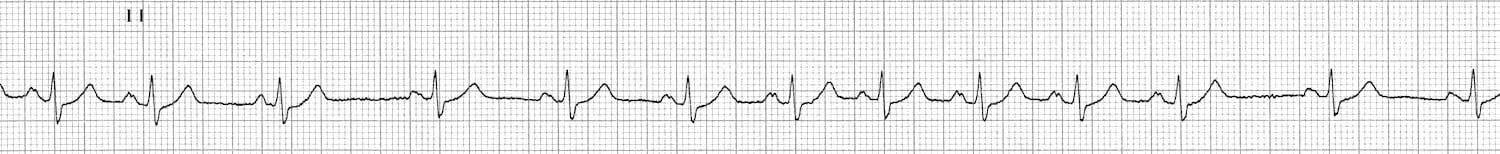
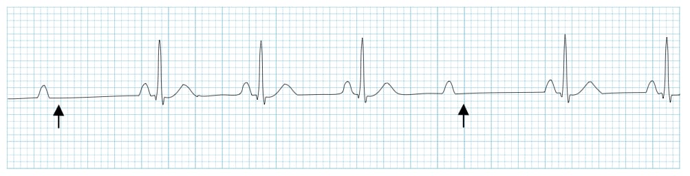
Here's an interesting example:
ECG strip with a perfectly-regular narrow-complex rhythm and a background of regular atrial tachycardia and irregular PR intervals

This is a lewis lead (increases amplitude of P-waves for easier rhythm identification) of a patient that I took care of after a mitral valve replacement. I initially ran this particular rhythm by several cardiologists and got several completely different identifications for this rhythm.

The atrial complexes are perfectly regular and monomorphic, and they appear to lack the "sawtooth" shape characteristic of atrial flutter. The R-R spacing is also perfectly regular, with a HR of 77. At first glance, this appears to be a simple case of atrial tachycardia with 4-1 conduction. But something unusual is going on here. Can you spot the anomaly?

Notice that the PR intervals are completely random. The P waves appear to have no relationship to the QRS complexes. This is not strange in-and-of-itself; there are plenty of irregular atrial arrhythmias that produce irregular PR intervals, but if the ventricles are following the atria, then an irregular impulse must produce an irregular rhythm, and if a regular impulse is producing a regular rhythm, then the PR intervals must be regular. The fact that the R-R intervals remain perfectly regular here, despite the PR intervals being all over the place, tells us that the P waves are not linked to the QRS complexes... which means that the ventricular rhythm is not listening to the atrial rhythm. Recall that the heart doesn't really have one rhythm; it has two (atrial and ventricular). Those two rhythms are usually in synchrony, but here is a case where that synchrony has been broken.

But if the rhythm in the ventricles here (the QRS complexes) is not coming from the atrium, where is it coming from? Well, the QRS complexes are narrow, which means it's not a ventricular rhythm. This is an accelerated junctional rhythm, with a background of atrial tachycardia.

Why aren't any of the atrial impulses getting through to the ventricles here? It's because this patient had a mitral valve replacement. The mitral valve is very close to the AV node. Inflammation of the AV node can impair it, resulting in an AV node that is easily fatigued. When this patient entered an atrial tachycardia, those rapid impulses from the atrium were simply too much for the patient's inflamed and tired AV node, and the AV node stopped transmitting the impulses. At that point the conduction system tissue a little farther down the track, either in the AV node or somewhere in the bundle of His, lacking any input from the atria and irritated by the inflammation from the surgery, produced an accelerated escape rhythm.

Now, I know what you're thinking: "Zero conduction through the AV node? That sounds like a 3° AV block to me." That's certainly what I thought, but the electrophysiologists that I spoke to regarding this rhythm informed me that complete blockage of the AV node that is due to fatigue caused by rapid atrial impulses is not a 3° AV block. Unfortunately they were unable to offer me an alternative definition for this manner of transient complete AV block (perhaps such a circumstance is rare enough that there is no commonly-accepted definition). I'm no electrophysiologist so I yield to their authority, but suffice to say, this is functionally identical to a 3° AV block.

In case you're wondering how we knew this block was rate-dependent, I actually watched this patient transition multiple times between this rhythm and sinus rhythm with a 2° type-1 AV block. Whenever the patient's atria exited Afib and returned to NSR, the AV node would resume transmission of impulses and the atrium would recapture the ventricles.

One big takaway here is that AV blocks can be rate-dependent just like bundle branch blocks. This is also a perfect example of how simple definitions and memorization of ECG patterns in textbooks can fail you, and how a basic understanding of the mechanics of the conduction system can elucidate what is happening in a patient's heart and the clinical implications, even in edge cases where professionals are unable to agree on a definition for what's happening!

To summarize some important rules for identifying junctional rhythms:

Ventricular Rhythms

Ventricular rhythms are always wide-complex, because they originiate in the ventricular myocardial tissue and don't use the efficient highway of the conduction system to spread across the heart. Without that highway system, it takes much longer for the depolarization to spread across the heart (like driving off-road), hence the wider QRS complex. Ventricular rhythms can sometimes produce retrograde P-waves that appear after or buried inside the QRS, often inverted in the inferior leads, just like the inverted P-waves produced by junctional rhythms. This is because the impulse from the ventricles can travel backwards up the conduction system into the atria, depolarizing the atria and producing a P-wave after the QRS. Ventricular rhythms will never produce a P-wave before the QRS complex, because that only happens when the depolarization impulse reaches the atrium before the ventricles, but the depolarization impulse of a ventricular rhythm starts in the ventricles by definition, so it's impossible for the P-wave to appear before the QRS in a ventricular rhythm (unless there is a 3° block and the atrium is producing its own P-waves with no relationship to the QRS complexes). Ventricular rhythms are often easily identified by their combination of slow rate (less than 40), wide complexes, and lack of relationship to P-waves if there is a 3rd degree block present and a background atrial rhythm.

Here's an example of a patient transitioning back and forth between NSR and an accelerated idioventricular escape rhythm:
ECG strip showing an accelerated idioventricular rhythm

Remember, not all junctional rhythms and ventricular rhythms are the result of blocks or sinus pauses. Sometimes for one reason or another the junction or ventricles will simply start going faster than the sinus node. Since the heart's tissues will respond to the first impulse they receive and then become refractory to subsequent impulses for a little while, the heart will follow the fastest pacemaker. The above case of a patient transitioning back and forth between sinus rhythm and a ventricular rhythm could be the result of the ventricles taking over during sinus pauses, or it could simply be the result of a sinus arrhythmia where the ventricles are taking over ("escaping") every time the sinus node slows down too much.

You can think of the hearts various pacemakers as being in a race to "capture" the rhythm of the heart. This analogy of the heart's pacemakers racing each other is the origin of much electrophysiological jargon. For example the terms "capture" and "escape" are a reference to this idea of a race for control of the rhythm. When a beat is dropped in a 2° AV block for example, a junctional or ventricular pacemaker may "escape," perhaps producing a single beat, or multiple beats before the atrium "recaptures." Sometimes a small pause is all a junctional or ventricular escape rhythm needs to capture the heart for many beats even if the escape rhythm is slightly slower than the atrial rhythm. This is because of something called "retrograde suppression" of the atrium. Each beat from the junction or ventricles will travel backwards up the conduction system through the AV node, at the same time as impulses are coming down from the atrium. These impulses collide with each other and cancel each other out, like two stubborn goats crossing different ways on a log. As long as this is happening the atrium is unable to get an impulse through to recapture the ventricles from the junction, even if the atrium is slightly faster than the junction, until such a time that the atrium sends an impulse at just the right moment to avoid the upwards traffic and recapture the junction and ventricles.

1° Blocks

Everybody knows what a 1° AV block looks like, but as we explore the differences between 1st, 2nd, and 3° blocks let's really think about what's happening in the conduction system and why these blocks manifest as they do on an ECG strip. A 1°AV block is a manifestation of AV node impairment or fatigue. Some degree of conduction fatigue when exposed to rapid signals is actually normal and healthy; a feature and not a bug of the heart's conduction system. Normal fatigue helps prevent the conduction system from transmitting overly-rapid signals from your atrium to your ventricles. Allowing too many impulses through too quickly could be deadly, so it's a good thing that our conduction systems are a little bit lazy and refuse to run too many signals through too quickly.

That said, conduction systems can become more fatigued than normal, and begin transmitting impulses more slowly, and eventually falling behind and dropping some of the impulses entirely. The AV node is part of the conduction system, and if the AV node becomes fatigued it may manifest a 1° AV block, and as it becomes more fatigued that 1° AV block may progress to a 2° AV block, and then to a 3° AV block.

ECG strip showing normal sinus rhythm with a long PR interval and P-waves buried in the T-waves

The above rhythm may appear at first glance to be an accelerated junctional rhythm, but notice the P-waves buried in the tail-end of the T-waves. The PR intervals are wide but they are regular. This is a very tired AV node doing its best to transmit all of the atrial impulses, and barely managing it by the looks of it.

2° Blocks

As the AV node becomes more fatigued, a 1° AV block may progress to a 2° type-1 AV block. But let's not just memorize that fact. Let's understand why! At this point the tired AV node can no longer transmit every impulse from the atria, and it starts dropping some of them. But when it drops an impulse, this actually gives the AV node a much-needed rest, and when the next impulse comes along from the atrium, the AV node is well-rested and transmits the signal much more quickly, resulting in a shorter PR interval. As the next impulses arrive, the AV node once again becomes progressively more fatigued until it gives up and drops a signal again. This once again gives the AV node some much-needed rest, shortening the PR interval and starting the cycle all over again.

Now that you know why 2° type-1 blocks look the way they do, look upon this example with your new eyes of wisdom! Don't just see a pattern of lines... see the conduction system! See the tired AV node working hard and intermittently failing to deliever those impulses to the ventricles.

ECG strip showing a Mobitz-1 AV Block

It's like the chocolate factory episode of I love Lucy. Imagine sinus node impulses are the pieces of chocolate, and the AV node is Lucy trying her best to wrap them and send them down to the ventricles...but she's not quite able to get all of them wrapped in time.

As for Mobitz-II, electrophysiological explanations of why there are intermittent dropped beats without any prolongation of the PR interval are beyond the scope of this page. Some literature states that 2° type-2 blocks tend to be physically farther below the AV node and closer to the bundle of His than 2° type-1 blocks, and fatigue of the conduction system below the AV node does not produce transduction delays the same way fatigue of the AV node itself does. Rather, it simply produces intermittent failures of transduction. An example of this is a bilateral bundle branch block. If one of the bundle branches is fully blocked, and the other is fatigued and intermittently blocking impulses, this will produce a Mobitz-II pattern of "dropped beats" without prolongation of the PR interval.

Here's an example of a Mobitz-II AV block:
ECG strip showing a Mobitz-2 AV Block
3° Blocks

3° AV blocks occur when the AV node is no longer able to transmit any signals at all between the atria and the ventricles. It's important to recognize that this will always result in two distinct rhythms for the heart, the rhythm of the atria, and the rhythm of the ventricles (I'm including asystole in the definition of "rhythm" here). The atrium doesn't have to be in asystole or sinus rhythm, it can be in Afib, Aflutter, sinus tach, MAT, sinus arrhythmia, ectopic bradycardia, you name it. The atrium can do whatever it wants to in a 3° block, and the ventricles are none-the-wiser. The ventricles will either adopt a junctional rhythm if there is a junctional escape, or they might initiate a ventricular escape rhythm if no junctional escape rhythm presents itself. Alternatively if no pacemaker below the block presents itself, the ventricles could sit in asystole while the atrium continues along in whatever rhythm it happens to be in.

The following is the classic example of a 3° block:
ECG strip showing a 3° AV block with a junctional escape

The narrow complexes tell us that this must be a junctional escape since we already know that it's not coming from the atrium due to the 3° block, and ventricular rhythms are always wide-complex. What if the complexes were wide? What would that tell us? Nothing! Remember, narrow complexes tell you that a rhythm is not coming from the ventricles, but a wide QRS complex can originate anywhere.